Craniotomy for Removal of a Brain Tumour
A craniotomy is a surgical procedure used to access the brain and remove a tumour. Surgery may be recommended to establish a diagnosis, relieve symptoms caused by pressure on the brain, or remove as much of the tumour as safely possible.
Understanding the Procedure
What is a Craniotomy?
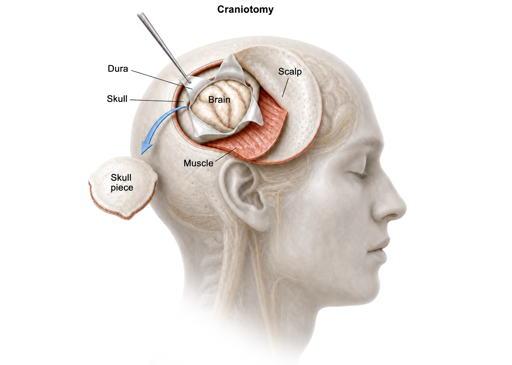
A craniotomy is a surgical procedure in which a small section of the skull is temporarily removed to allow access to the brain.
Once the tumour has been identified, specialised microsurgical techniques are used to remove all or part of the tumour while protecting the surrounding brain tissue. At the end of the procedure, the bone is replaced and secured using small titanium plates and screws.
A craniotomy is one of the most common procedures used to treat brain tumours.
Understanding Brain Tumours
Brain tumours can arise from the brain itself (primary brain tumours) or spread to the brain from cancers elsewhere in the body (secondary or metastatic tumours).
Common brain tumours include:
- Meningiomas — usually benign tumours arising from the lining surrounding the brain.
- Gliomas — tumours arising from cells within the brain itself, ranging from low-grade to more aggressive forms.
- Metastatic tumours — cancers that have spread to the brain from another part of the body.
Some tumours are benign (non-cancerous), while others are malignant (cancerous). Even benign tumours may require treatment if they are growing, causing symptoms, or placing pressure on important parts of the brain.
The goals of surgery vary depending on the type and location of the tumour. Surgery may be performed to:
- Confirm the diagnosis
- Remove as much of the tumour as safely possible
- Relieve pressure on the brain
- Improve neurological symptoms
- Assist planning of further treatment such as radiotherapy or chemotherapy
Not all brain tumours require complete removal, and in some situations preserving neurological function is more important than aggressive tumour removal.
Indications
When is this Procedure Recommended?
A craniotomy may be recommended when a brain tumour is causing symptoms or when tissue is required to establish a diagnosis.
Common reasons include:
- Meningioma
- Glioma
- Metastatic brain tumour
- Progressive tumour growth
- Headaches caused by increased pressure within the skull
- Seizures
- Weakness or numbness
- Speech difficulties
- Visual disturbance
- Balance or coordination problems
The Procedure
What Happens During Surgery?
- The head is carefully positioned and prepared for surgery.
- An incision is made in the scalp.
- A section of skull (bone flap) is temporarily removed.
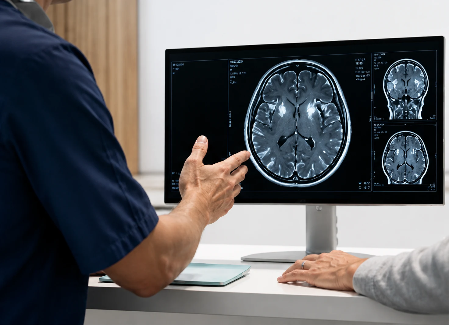
- The tumour is identified using microsurgical techniques and stereotactic image guidance, allowing the surgeon to precisely navigate within the brain and minimise disruption to surrounding structures.
- The tumour is removed either completely or partially, depending on its location and relationship to surrounding brain structures.
- The bone flap is replaced and secured.
- The wound is closed and a dressing is applied.
- Most procedures take approximately 2–6 hours, depending on the complexity of the tumour.
Related Conditions
Benefits
Potential Benefits
The goal of surgery is to safely remove tumour tissue while preserving neurological function.
Potential benefits include:
- Relief of pressure on the brain
- Improvement in neurological symptoms
- Reduction in headaches
- Improved quality of life
- Establishment of a definitive diagnosis
- Facilitation of further treatment if required
Recovery varies between individuals and depends on the type, size, and location of the tumour, as well as the extent of surgery performed.
After the procedure
Recovery After Surgery
Most patients spend several days in hospital following surgery. The exact length of stay depends on the complexity of the procedure and the speed of recovery.
Headaches, scalp discomfort, and fatigue are common during the early recovery period. These symptoms generally improve over the first few weeks.
Walking and gentle activity are encouraged as soon as it is safe to do so. Recovery should progress gradually and strenuous activity should be avoided until cleared by your surgeon.
Some patients benefit from physiotherapy, occupational therapy, speech therapy, or other rehabilitation services depending on the symptoms present before and after surgery.
Restrictions on driving vary depending on the underlying diagnosis, neurological symptoms, and whether seizures have occurred. Your surgeon will provide individual advice.
Return to work depends on the type of work performed, the nature of the tumour, and the extent of surgery. Recovery commonly takes several weeks and sometimes longer.
Follow-up appointments are usually scheduled at 2 weeks and 6 weeks after surgery. Results of tumour analysis and any additional treatment recommendations are discussed during follow-up.
Potential Complications
Risks and Complications
All surgery carries risks. Potential complications include:
- Infection
- Bleeding
- Blood clots
- Anaesthetic complications
- Seizures
- Cerebrospinal fluid leak
- Temporary or permanent neurological deficits
- Weakness or numbness
- Speech difficulties
- Visual disturbance
- Brain swelling
- Need for further surgery
Your surgeon will discuss risks relevant to your individual circumstances, including risks that relate specifically to the location of the tumour.
Seeking Advice About a Brain Tumour?
Our team can help explain your diagnosis, discuss treatment options, and determine whether surgery is appropriate.