Stereotactic Brain Biopsy
A stereotactic brain biopsy is a minimally invasive procedure used to obtain a tissue sample from a brain lesion or tumour. The sample is analysed by a pathologist to establish a diagnosis and guide further treatment.
Understanding the Procedure
What is a Stereotactic Brain Biopsy?
A stereotactic brain biopsy is a procedure used to obtain a small sample of tissue from an abnormal area within the brain.
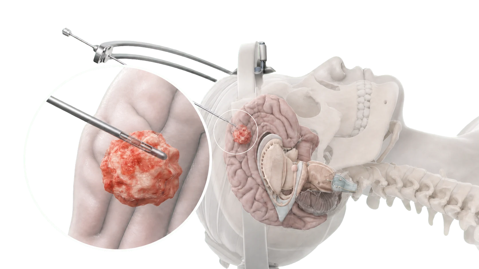
Using advanced imaging and stereotactic navigation technology, the surgeon can accurately target the lesion through a small opening in the skull. The tissue sample is then examined under a microscope to determine the nature of the lesion.
A stereotactic biopsy is often performed when a diagnosis cannot be established from imaging alone or when a tumour is located in an area where complete surgical removal may not be appropriate.
Understanding the Need for A Biopsy
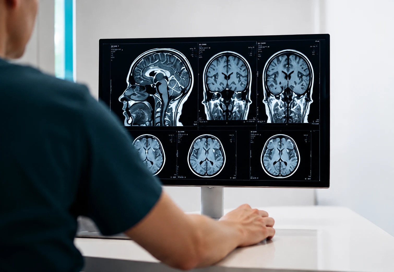
Brain imaging can often identify the presence of an abnormality, but scans alone cannot always determine exactly what it is.
A tissue diagnosis may be required to distinguish between:
- Glioma
- Lymphoma
- Metastatic tumour
- Inflammatory conditions
- Infection
- Other rare brain lesions
The information obtained from a biopsy helps guide decisions regarding surgery, radiotherapy, chemotherapy, observation, or other treatments.
In many cases, obtaining a diagnosis is the most important first step in developing an appropriate treatment plan.
Indications
When is this Procedure Recommended?
A stereotactic brain biopsy may be recommended when a brain lesion requires tissue diagnosis before treatment decisions can be made.
Common reasons include:
- Newly diagnosed brain lesion
- Suspected glioma
- Suspected lymphoma
- Suspected metastatic tumour
- Deep-seated brain lesions
- Lesions located near critical brain structures
- Uncertain diagnosis on MRI or CT imaging
The Procedure
What Happens During Surgery?
- Detailed MRI or CT imaging is used to plan the procedure.
- The head is secured and stereotactic navigation is established.
- A small incision is made in the scalp.
- A small opening is created in the skull.
- A biopsy needle is carefully guided to the target using stereotactic image guidance.
- Small tissue samples are obtained.
- The wound is closed and a dressing is applied.
- Most procedures take approximately 1–2 hours.
Related Conditions
Benefits
Potential Benefits
The goal of surgery is to establish an accurate diagnosis while minimising disruption to surrounding brain tissue.
Potential benefits include:
- Definitive tissue diagnosis
- Guidance for treatment planning
- Avoidance of unnecessary treatment
- Identification of tumour type and grade
- Improved decision-making regarding surgery, radiotherapy, or chemotherapy
Recovery varies between individuals but is generally quicker than after larger brain operations.
After the procedure
Recovery After Surgery
Most patients spend 1–2 nights in hospital following surgery for observation and monitoring.
Some mild headache or discomfort around the incision site is common and usually improves over the first few days.
Walking and light activity are encouraged soon after surgery. Most patients can gradually return to normal daily activities over the following weeks.
Driving restrictions vary depending on the underlying condition, neurological symptoms, and whether seizures have occurred. Your surgeon will provide individual advice.
Many patients are able to return to office-based work within 1–3 weeks depending on their recovery and underlying diagnosis.
Follow-up appointments are usually scheduled within 1–2 weeks. Biopsy results are reviewed and further treatment recommendations discussed.
Potential Complications
Risks and Complications
All surgery carries risks. Potential complications include:
- Infection
- Bleeding
- Blood clots
- Anaesthetic complications
- Bleeding within the brain
- Seizures
- Temporary or permanent neurological deficits
- Weakness or numbness
- Speech difficulties
- Inconclusive biopsy requiring further investigation
Your surgeon will discuss risks relevant to your individual circumstances, including risks that relate specifically to the location of the lesion.
Seeking Advice About a Brain Lesion or Tumour?
Our team can help explain your diagnosis, discuss treatment options, and determine whether a biopsy is appropriate.